- Premier Competition for High School Students Culminates at BIO International Convention
- Winner, Emily Wang, Developed New Fluorescent Proteins to Improve Biosensing
- Her Grandmother’s Battle with Cancer Inspired this Award Winning Research
I don’t know about your science achievements while in high school, but mine were limited to getting up early on Saturdays to go to Biology Club to dissect worm, starfish and cat specimens to study anatomy—and trying not to pass out from noxious formaldehyde preservative! Thus, I am constantly amazed by the level of complexity and maturity that I see in young science students today, and I always look forward to seeing who will win the annual BioGENEius Challenge.
The BioGENEius Challenge is the premier competition for high school students that recognizes outstanding research in biotechnology. The Challenge is organized by the Biotechnology Institute, a U.S. based nonprofit organization dedicated to biotechnology education. Its mission is to engage, excite and educate the public, particularly students and teachers, about biotechnology and its immense potential for solving human health, food and environmental problems.
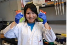 Emily Wang proudly showing her International 2014 BioGENEius Challenge award winning fluorescent proteins. Photo credit: Emily Wang.
This past June, Emily Wang—a graduating senior at Gunn High School in Palo Alto, California—was named the winner of the 2014 International BioGENEius Challenge. A panel of judges found that Emily’s research in developing fluorescent proteins to improve biosensing helped her stand out from the 14 other finalists from across the U.S. and Canada competing for the Challenge's top prize, a $7,500 cash award.
Emily’s win was announced during a luncheon at the hugely popular 2014 BIO International Convention, and was keynoted by even more famous Sir Richard Branson. The International BioGENEius Challenge is one of the few international competitions to host participants at a leading industry conference, thereby providing students with unparalleled access to companies, scientists and innovators in biotechnology.
Emily was not only evaluated on the quality of her research, but also on her research poster presentation and responses to questions testing her scientific knowledge. Moreover, each student’s research was reviewed for the potential commercial and practical applications of their project.
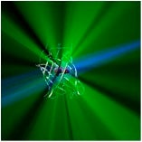 Computer artwork of the molecular structure of green fluorescent protein (GFP). Some central atoms are represented as spheres. The molecule has a cylindrical structure formed from beta sheets (ribbons). GFP is found in the Pacific jellyfish (Aequorea victoria). It fluoresces green when blue light is shone on it, as depicted here (taken from fineartamerica.com via Bing Images).
Emily’s project, titled, “Illuminating Disease Pathways: Developing Bright Fluorescent Proteins to Improve FRET Biosensing,” seeks to help visualize disease pathways, including cancer metastases.
Before I get into the details of Emily’s project, I should pause to mention the second, third, and fourth place winners, who won $5,000, $2,500 and $1,000, respectively. Those winners were:
- Logan Collins, Fairview High School, Boulder, CO
- Neil Davey, Montgomery Blair High School, Silver Spring, MD
- Nathan Han, Boston Latin School, Boston, MA
Emily’s Project for the International 2014 BioGENEius
With the help of Gayle Kansagor of the Biotechnology Institute, I contacted Emily to get more specific information about her award winning research project for this blog post, and inquire about the availability of the fluorescent proteins that she developed. Emily kindly provided me with the following first-person account.
Layman’s Description
I aimed to create a tool to visualize diseases at the molecular level. I developed Clover3, a bright green fluorescent protein, and mRuby3, a bright red fluorescent protein. Clover3-mRuby3 can be used to image neurons to investigate Alzheimer’s disease, detect and track cancer growth, and observe biological activities with greater clarity than before.
Abstract
The discovery and development of fluorescent proteins, recognized by the 2008 Nobel Prize in Chemistry, enabled a revolution in biological microscopy and sensing. Biosensors employing fluorescence resonance energy transfer (FRET) between fluorescent proteins are powerful tools to non-invasively report biochemical events within living cells. The development of new FRET sensors, however, remains difficult, often due to low FRET dynamic range, which worsens detection of subtle or transient cellular responses. I engineered a new green fluorescent protein Clover3, which confers increased FRET dynamic range onto biosensors and shows improved quantum yield. I also developed a new red fluorescent protein mRuby3, which improves extinction coefficient. Clover3-mRuby3 may benefit a variety of biomedical applications, including imaging of neural structures to investigate Alzheimer’s disease, detecting and tracking of cancer metastases, and monitoring signaling pathways to elucidate disease mechanisms. Clover3-mRuby3 may be used to visualize biological activities with greater clarity than before, which may advance current understanding of illnesses to create new therapies and medicine to improve human health.
Motivation
When I was little, my paternal grandmother passed away due to Stage IV brain cancer. The late detection of cancer filled me with indignation, and I longed to understand how cancer furtively grew and spread. I wanted to create a biological imaging tool, a bright fluorescent protein, to help researchers visualize cancer activities at the molecular level. After reading countless medical papers about fluorescent proteins, I became convinced that the future of cancer research lies in creating new imaging tools from fluorescent proteins. I learned that the innovative field of fluorescent protein engineering had potential to revolutionize cancer research. I also learned, however, that current fluorescent protein technology still had limitations, such as poor photostability and low fluorescence resonance energy transfer (FRET). I wanted to improve current fluorescent proteins and develop a new one that could serve as a valuable tool for scientists to understand disease pathways. With my interest in fluorescent proteins, I contacted Professor Michael Lin at Stanford University's Department of Bioengineering, which was how I started my current project.
Publication Potential for and Availability of Emily’s Proteins
I asked Emily about the availability of the new fluorescent proteins for research use and she said that one of the earlier proteins, Clover2 can be found on AddGene. Not having heard about AddGene, I visited its website and learned that Addgene is a non-profit plasmid repository which is “dedicated to helping scientists around the world share high-quality plasmids.” Moreover, it has been amazingly effective—in my opinion—at achieving this goal: over the past ten years Addgene has shipped over 350,000 individual plasmids to 5,000 different research institutions. If you’re interested in knowing which of these are the “Top 10” plasmid technologies that have been distributed, you can consult this list.
When I asked Emily about whether this work was being published, she said that it was going to be. However, as of writing this post, I couldn’t find a citation in PubMed, so we’ll have to wait for that publication. I also inquired about a patent application, and was told by her that this was in the process of being dealt with.
Concluding Comments
If you’re wondering, as I was, where Emily has gone for further education, I connected with her on LinkedIn, and learned that she’s a student at Harvard University—undoubtedly studying hard and looking forward to doing more great research.
Emily’s research is a prime example of how sophisticated high school science has become, partly due to increased awareness of the importance of biotechnology in all aspects of modern society.
BTW, in contrast to Emily’s DNA-based plasmid delivery of encoded biosensor proteins, I find it interesting and exciting that modified mRNA is now being used as an alternative approach that provides various advantages. TriLink has recognized the importance of this new methodology, and now offers modified mRNAs encoding a wide variety of reporter proteins that include several popular “colors” of fluorescent proteins. You can find out more about these and other mRNA products on the TriLink website.
As always, your comments are welcomed.
Postscript
As mentioned in Emily’s abstract above, the importance of green fluorescent proteins (GFPs) was recognized by the 2008 Nobel Prize in chemistry, which was jointly awarded to Osamu Shimomura of Boston University and Marine Biological Laboratory, Martin Chalfie of Columbia University, Roger Y. Tsien at the Department of Chemistry and Biochemistry, University of California, San Diego.

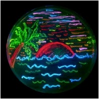 The diversity of genetic mutations is illustrated by this San Diego beach scene drawn with living bacteria expressing 8 different colors of fluorescent proteins derived from GFP and dsRed. Taken from Wikipedia.
Because of the transformative utility of GFPs and modified analogs having different colors as labels, there are many versions with different excitation and emission properties to suite specific requirements. I couldn’t resist including this cute illustration of spectral diversity provided by the Tsien Laboratory in sunny San Diego.