- Discovered 30 years ago, exosomes are now implicated in cell-to-cell communication
- Exosome-based commercialization is on the rise
- NIH is also pouring more money into exosome research
Exosomes aren’t cellular "garbage bags" after all
Previous postings here highlighted 30th anniversaries of PCR discovery and automating oligo synthesis, which both occurred in 1983. Remarkably, in the same year, two research groups independently reported—within one week of each other—that, in reticulocytes (immature red blood cells), transferrin receptors associated with tiny ~30-100nm diameter vesicles are literally jettisoned from maturing blood reticulocytes into the extracellular space—but not as “garbage bags.” The name “exosome” for these extracellular vesicles was coined several years later to differentiate them from traditional “endosome” compartments inside cells. Readers interested in the context of these parallel and pivotal observations, as well as an overview of key follow-on reports, are referred to a first-hand account by Harding, Heuser & Stahl. Therein, it was simply stated that “[e>
xosomes have become relevant to many fields.” I quantified this scientific pervasiveness by searching PubMed for exosome(s) or exosomal, which gave ~2,300 references since 1983, with the vast majority of these appearing during the past ten years and showing significant year-over-year frequency. During 2012, these publications appeared at an average of one per day! Not surprisingly, this groundswell of scientific interest led to founding the International Society for Extracellular Vesicles, as well as the American Society for Exosomes and Microvesicles. Among relatively recent reviews of exosomes that can be easily found in PubMed, the short account by Raposo & Stoorvogel published this year in J. Cell Biol. provides an easy to read introductory overview, especially with regards to exosomal RNA (exRNA)-mediated intercellular communication.
"Cell speak" - so that’s how they do it!
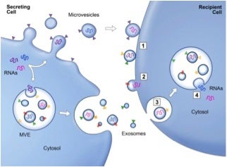
If you ever wondered how a cell communicates with other cells (whether like or unlike), multiple processes are involved, some of which include exosomes. Fusion of the exosomes with the plasma membrane of the recipient cell, as depicted below, allows for transfer of the internal components to the target cell and thus, the transfer of information. The molecules being transferred can include proteins, such as receptors or enzymes, and RNA that are collectively referred to as exRNA. As you can see in the schematic, membrane-associated (triangles) and transmembrane proteins (rectangles) and RNAs (curved symbols) are selectively incorporated into multivesicular endosomes (MVEs) or into microvesicles (MVs) budding from the plasma membrane. MVEs fuse with the plasma membrane to release exosomes into the extracellular milieu. MVs and exosomes may dock at the plasma membrane of a target cell (1). Bound vesicles may either fuse directly with the plasma membrane (2) or be endocytosed (3). Endocytosed vesicles may then fuse with an endocytic compartment (4). Both pathways result in the delivery of proteins and RNA into the membrane or cytosol of the target cell. Fusion and endocytosis are only represented for exosomal vesicles, but plasma membrane–derived MVs may have similar fates. The ability to influence gene expression in distant cells through exosomes presents a remarkable model for cell-to-cell signaling that offers an entirely new perspective on intercellular communication. This also has potential therapeutic applications, such as in diagnosis, intervention, and artificial gene/mRNA delivery.
Protein and RNA transfer by extracellular vesicles. Copyrighted by Raposo & Stoorvogel and published by Rockefeller University Press.
In a recent, brief review of this subject, cell communication is classified broadly as contact-dependent, paracrine, synaptic, or endocrine. Traditional cell-cell communication occurs by several means, including chemical receptor-medicated events (e.g., chemotaxis), direct cell-cell contact, and cell-cell synapses. Contact-dependent cellular communication involves a physical connection between the participating cells. Contact-independent communication can occur between cells not directly linked, and may be mediated through paracrine or endocrine mechanisms involving the extracellular transfer of information molecules (e.g. hormones).
In 2007 Valadi et al. reported that exosomes from mast cells contained mRNA from ~1,300 genes, many of which were not present in the cytoplasm of the donor cell. In vitro translation proved that the exosome mRNAs were functional, and analysis of total RNA derived from exosomes revealed the presence of small RNAs, including miRNAs. Moreover, the RNA from mast cell exosomes was transferable to other mast cells. The Valadi publication, which sparked intense interest in this novel mechanism of cell-cell communication, has been cited more than 1,300-times—an amazing numerical coincidence!—especially in the molecular biology of cancer.
Placental exosome-mediated immune protection of the fetus: feeling groovy in a cloud of exosomes
This section heading is actually the title of a fascinating article by Lucia Mincheva-Nilsson published in Expert Review of Obstetrics & Gynecology in 2010. The abstract states that “[r>
ecently, it was discovered that the syncytiotrophoblast of human placenta continuously and constitutively secretes exosomes throughout pregnancy. These exosomes, delivered directly in the maternal blood surrounding the chorionic villi of the placenta, are immunosuppressive and pluripotent carrying proteins, mRNA and miRNA that can influence a number of biologic mechanisms and promote the fetal allograft survival. The current knowledge regarding placental exosomes and their role in pregnancy is summarized and discussed in this article.” A publication by Ng et al. in PloS ONE in 2013 entitled Endometrial Exosomes/Microvesicles in the Uterine Microenvironment: A New Paradigm for Embryo-Endometrial Cross Talk at Implantation can be accessed here. These investigators report that 13 of the 227 miRNA were specific to exosomes/MVs, while a further 5 were not present in these. The most abundant miRNA in exosomes/MVs were hsa-miR-200c, hsa-miR-17 and hsa-miR-106a. Bioinformatic analysis showed that the exosome/MV-specific miRNAs have potential targets in biological pathways highly relevant for embryo implantation.
Tumor exosomes manipulate cancerous growth and invasiveness
In a 2013 publication in Neoplasia, Zöller and coworkers note that one important factor in tumor biology is the capacity of tumor cells to create a micro-environment by recruiting and modulating non-transformed cells that favor tumor cell survival and spreading/propagation. They cite considerable previous experimentation demonstrating that exosomes are abundantly delivered by tumor cells, and are known to constitutively express adhesion molecules that contribute to exosome-selective target-cell binding. In addition, exosomes binding can induce target cell activation, and through transfer of exRNA, contribute to target cell modulation. These exosome activities were said to be of likely importance in cross talk between cancer stem cell exosomes and neighboring cancer cells, although details are lacking. Finally, in the aforementioned citation, evidence is provided that indicated tumor exosome-mediated degradation of the host matrix. This strongly supports host-cell migration and invasiveness, as well as host-cell proliferation and apoptosis resistance. These authors conclude that the impact of tumor exosomes on the host matrix can have severe consequences on tumor progression.
Kucharzewska et al., in Proc. Natl. Acad. Sci. in 2013, emphasize that hypoxia, or low oxygen tension, is a major regulator of tumor development and aggressiveness. However, how cancer cells adapt to hypoxia and communicate with their surrounding microenvironment during tumor development remain important questions. Therein they show that secreted vesicles with exosome characteristics mediate hypoxia-dependent intercellular signaling of the highly malignant brain tumor glioblastoma multiforme (GBM). In vitro hypoxia experiments with glioma cells and studies with patient materials reveal the enrichment in exosomes of hypoxia-regulated mRNAs and proteins (e.g., matrix metalloproteinases, IL-8, PDGFs, caveolin 1, and lysyl oxidase), several of which were associated with poor glioma patient prognosis. Importantly, they concluded that “exosomes constitute a potentially targetable mediator of hypoxia-driven tumor development, and that the exosomal molecular signature may serve as a noninvasive biomarker to assess the oxygenation status and aggressiveness of malignant tumors.”
Xaio et al., in PloS One in 2012, reported similar conclusions for melanoma-derived exosomes that were found to have unique gene expression signatures, miRNA and proteomics profiles compared to exosomes from normal melanocytes. These conculsions were based upon their first-ever in-depth screening of whole transcriptome/miRNome/proteome expression in melanoma exosomes. They concluded that these and future results “define targets that can be translated into clinical applications as non-invasive biomarkers or as therapeutic targets for melanoma patients.” As would be expected, this and other reports of possible novel, exosome-related drug-targeting strategies and biomarkers has fueled considerable commercial interest.
Exosome-Based Commercialization
Tools
Basic and applied health-related science “tool” providers have been quick to commercialize a wide array of exosome-based products. Not surprisingly, early offerings included various “sample prep” products for enrichment or isolation of exosomes that, because of their relatively small size, is nontrivial compared to other types of sample prep. BioCat, Invitrogen (Life Technologies), Norgen Bio, and System Biosciences are some of the suppliers of exome-based products.
Among methods for subsequent analysis of exRNAs, traditional qPCR and—especially—various library preparations for major next-generation sequencing platforms are available, such as those from Life Technologies for Ion Torrent sequencing.
Diagnostics
Higher “valued added” applications utilizing exRNA data include diagnostics. Next-generation sequencing of exRNA is being applied to identify potential biomarkers, as exemplified in a recent report by Kurochkin and co-workers entitled, who sequenced exRNA from breast cancer cell lines, and concluded that their “data revealed that exosomal transcripts are representative of their cells of origin and thus could form basis for detection of tumor specific markers.”
An up-to-date indication of many of the players and hot topics in exosome research can be found at the website for the inaugural Exosomes and Circulating Biomarkers 2013 Summit, recently held in San Diego. The summit focused on cutting-edge science with presentations from top-tier academic and industrial researchers. Exosome Diagnostics and Caris Life Sciences are two companies who participated in the summit and offer unique technologies/products based for the exome market.
Exosome Diagnostics, which is developing biofluid-based molecular diagnostic tests for use in personalized medicine, announced in June 2012 that it will use Applied Biosystems qPCR instrumentation for its exosome biofluid in vitro diagnostics oncology programs involving multi-center clinical trials in brain cancer and prostate cancer. In July 2013 Exosome Diagnostics announced a partnership with Qiagen to develop and commercialize high-performance, co-branded kit products for the capture and processing of RNA (and DNA) from biofluid exosomes and other MVs. The stated goal is to provide products enabling researchers and drug developers to obtain repeated, real-time genetic "snapshots" of disease from patients' blood, urine or cerebrospinal fluid without the need for tissue biopsy.
Caris Life Sciences, which services personalized medicine through advanced laboratory testing, including tumor profiling and innovative blood-based cancer diagnostics, has developed what it calls Carisome® Microvesicle Technology. This technology platform has the ability to identify and characterize circulating MVs released in the blood that serve as a signaling device for various types of cancer. Caris uses both DNA- and antibody-based assays to obtain circulating MV “biosignatures” that have been associated with colorectal, prostate, breast, and lung cancers, as described here in various posters, papers, and presentation abstracts.
Around the same time as the summit, Genome Web announced that Aethlon Medical (see below) launched its wholly owned subsidiary, Exosome Sciences, to "pursue exosome-based strategies" for diagnosing and monitoring the progression of cancer, infectious diseases, and other conditions. Exosome's research facility will be within a CLIA-certified laboratory in Langhorne, PA. Aethlon said it will contribute diagnostic-related technology to Exosome, including the Enzyme Linked Lectin Specific Assay, which has been validated to identify the presence of exosomes underlying HIV, tuberculosis, and cancers such as ovarian, melanoma, breast, lymphoma, and colorectal.
Therapeutics
Therapeutic strategies related to exosomes fall broadly into two categories: targeted delivery of therapeutic agents and intervention in exosome-mediated diseases. A review of the former—wittily entitled FedExosomes: Engineering Therapeutic Biological Nanoparticles that Truly Deliver —was published earlier this year by Marcus & Leonard in Pharmaceuticals (Basel). Therein they summarize recent progress in harnessing exosomes for therapeutic RNA delivery, discuss the potential for engineering exosomes to overcome delivery challenges and establish robust technology platforms, as well as describe both potential challenges and advantages of utilizing exosomes as RNA delivery vehicles.
Regarding interventional strategies, Gould et al. published (Proc. Natl. Acad. Sci.) a widely cited article entitled The Trojan exosome hypothesis wherein it was proposed that retroviruses exploit exosome exchange for both the biogenesis of retroviral particles and a low-efficiency but mechanistically important mode of infection. They state that “retroviruses pose an unsolvable paradox for adaptive immune responses, that retroviral antigen vaccines are unlikely to provide prophylactic protection, and that alloimmunity is a central component of antiretroviral immunity. Finally, the Trojan exosome hypothesis has important implications for the fight against HIV and AIDS, including how to develop new antiretroviral therapies, assess the risk of retroviral infection, and generate effective antiretroviral vaccines.”
A quite different and very recent example of possibly intervening in exosome function to fight disease comes from the surprise discovery by Melbourne scientists that malaria parasites can “talk” to each other—a social behavior to ensure the parasite's survival and improve its chances for being transmitted to other humans. In his account of this fascinating work in BioQuick News, Michael D. O'Neill notes that these findings “could provide a niche for developing antimalarial drugs and vaccines that prevent or treat the disease by cutting these communication networks.”
In terms of actual “productization,” Aethlon Medical in San Diego has developed the Aethlon Hemopurifier® medical device. The Hemopurifier® captures exosomes underlying cancer, including colorectal, lymphoma, melanoma, ovarian, and breast cancer. In collaboration with the Sarcoma Oncology Center based in Santa Monica, Aethlon is studying the ex vivo effectiveness of its Hemopurifier® to remove immunosuppressive exosomes from the blood of advanced-stage cancer patients. The study will evaluate 25 patients, including five patients with metastatic cancer of the following types; non-small cell lung cancer, prostate cancer, melanoma, head and neck cancer, and sarcoma.
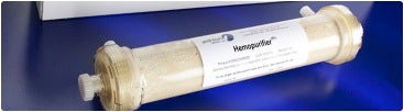 The Aethlon Hemopurifier® (taken from aethlonmedical.com).
A related medical device, HER2osome™, provides a therapeutic strategy to maximize the ability of the immune system and established drug therapies to combat HER2+ breast cancer, which is characterized by aggressive growth and poor prognosis resulting from the over-expression of HER2 protein. HER2osome™ is intended to inhibit HER2+ breast cancer progression by reducing the circulatory presence of HER2 protein and tumor-secreted exosomes that contribute to the development and progression of breast cancer. HER2osome™utilizes immobilization of a HER2 antibody and an exosome-targeted affinity agent to provide a mechanism to clear both targets from the circulatory system of HER2+ breast cancer patients. Published research indicates that breast cancer exosomes suppress the immune response, stimulate angiogenesis, contribute to the spread of metastasis, and inhibit the therapeutic benefit of Herceptin® (trastuzumab), a leading monoclonal antibody treatment against the HER2+ breast cancer. As an adjunct therapeutic candidate, HER2osome™ offers to fill an unmet medical need and enhance the benefit of Herceptin® and standard of care chemotherapies without adding drug toxicity or interaction risks.
NIH awards $17M for exRNA studies
This past August GenomeWeb News reported that the NIH has awarded $17 million to fund 24 new research projects seeking to understand how exRNA is involved in communication between cells, and how they may be used as biomarkers for diseases. This new funding will fuel efforts to examine exRNA biology and to develop tools for applying new discoveries about exRNA in research and in disease diagnosis and treatment.
James Anderson, Director of the Division of Program Coordination, Planning, and Strategic Initiatives is quoted as saying that, "[t>
o harness exRNA's enormous potential for diagnostics and therapeutics in a broad range of diseases, we first need to understand more about different types of exRNA, how cells make and release it, how it travels through the body, how it targets and affects specific cells, and how the amount and type of exRNA can change in disease."
More specifically, the National Center for Advancing Translational Sciences (NCATS) will administer 18 of the awards, including 10 projects seeking to develop biomarkers from exRNA, and 8 efforts aimed at designing new ways to use exRNA in treatments. The NCI will oversee 5 studies that will address how cells make and release exRNA, how and where it moves through fluids to other cells, and how it can change cellular function. The biomarker-focused projects overseen by NCATS will involve studies seeking to test out the clinical utility of using exRNA to develop biomarkers in a range of disease areas. For example, investigators at Brigham and Women's Hospital will study microRNAs in the blood of patients with multiple sclerosis to see if they can be used as markers for the disease or for diagnosing or monitoring its progression.
Also under the program, The National Institute on Drug Abuse has awarded a grant to Baylor College of Medicine to house the data generated through all of these exRNA studies, and to create a public exRNA Atlas website that will serve as a community resource for exRNA research standards, protocols, data tools, and technologies.
In another effort, researchers at Oregon Health and Science University will use a grant to try to find out if exRNA could potentially be used as a diagnostic biomarker for Alzheimer's disease that could enable the disease to be diagnosed earlier. Other projects will seek to find out if exRNA can be used as biomarkers for human glioma, gastric and hepatocellular cancer, placental dysfunction during pregnancy, and to predict outcomes after brain injury, among others.
For those of you who would like more information about exRNA and related industry news, you can find that here. If you wish to participate in conversations about exosomes, check out ExosomesTalk curated by scientists at Life Technologies.
I hope you find the same excitement that I do in the ever expanding and developing field of exome research. I’m very interested to hear what you may be doing in relation to this field, so as always, your comments are welcomed!
Postscript
exRNA Sneak Preview?
The many notable achievements of Paul Zamecnik include co-discovery to tRNA, and then pioneering work in antisense therapeutics. His seminal antisense publications in 1978 led—in a roundabout way—to discovering that cells contain relatively short (~5 to 50-nt) endogenous oligonucleotides, as reported in Proc. Natl. Acad. Sci. in 1987. When I read this paper and its discovery, the following commentary in it struck me as being very exciting—little did I know then that it foreshadowed future discoveries involving exRNAs!
 Paul Zamecnik (1912-2009) (taken from Bing Images)
“The results indicate that oligoribonucleotides exist free in eukaryotic cells. The amounts of cellular material at hand and the methods of separation used have not enabled us as yet to isolate pure oligonucleotides for sequence determination…”
“It is perhaps premature to discuss herein the possible function of the cellular oligonucleotides herein described. It is, however, worth considering that they may play a regulatory role in intracellular metabolism and may conceivably travel from one cell to another in a similar role.”
Click here for the classic paper on the discovery of tRNA by Zamecnik and coworkers.
Sad Endnote
During preparation of this blog, a NY Times obituary reported that Anthony Pawson, who identified the specific protein interactions involved in cell signalling, died on August 7th at the age of 60.
Scientists had long known that cells communicated, but no one knew the exact cellular mechanism involved until Dr. Pawson’s research pinpointed it: a protein structure on the surface of every cell membrane. The structure, which he called the SH2 domain, serves as a landing pad for signaling proteins, which in turn set off a molecular chain reaction carrying information to the cell’s nucleus.
 Anthony Pawson in 2009; his discoveries helped spur the development of drugs for cancer, diabetes and other diseases. (Photo taken from nytimes.com)
According to the NY Times, Anthony Hunter, a professor of molecular and cell biology at the Salk Institute Cancer Center in San Diego, called the identification of the SH2 domain an “enormously influential idea” that introduced scientists to a fundamentally new principle about how cells work.
“It was a seminal finding,” said Dr. Hunter, who collaborated with Dr. Pawson on several papers about cell communication.
Dr. Pawson’s research opened a new field of study into the causes and effects of breakdowns in cellular communication. Studies based wholly or in part on his discoveries have produced new treatments for cancer, autoimmune diseases, diabetes and heart ailments, essentially by blocking or unraveling intercellular miscues. Perhaps the best-known of these is Gleevec, a cancer drug that blocks the abnormal cell signal that causes a rare form of blood cancer called chronic myelogenous leukemia.


