- Human brain connectome: the last frontier?
- Is the UK making a real HAL 9000?
- MRI imaging of love and other emotions
To be perfectly honest, writing a blog about USA and UK big-buck backed ‘brain initiatives’ was on my mental—pun intended—‘to do’ list for some time, but I couldn’t see any direct relevance to trends in nucleic acids research. I changed my mind—pun intended—and opted to write this post after a recent publication in highly respected PNAS grabbed my attention. The publication dealt with brains and sex—and since Valentine’s Day is just around the corner, the time seemed right.
In this publication, brains were shown to be ‘hard wired’ very differently based on sex, and—more interestingly—these so-called ‘structural connectomes’ were associated with different behavioral characteristics. Male brains are structured to facilitate perception and coordinated action, whereas female brains are designed to facilitate communication between analytical and intuitive processing modes. My first reaction was that these ‘connectome’-derived behaviors aligned with males being ‘action’-oriented and females having ‘women’s intuition’.
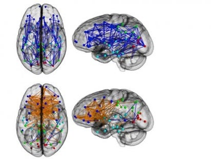 Brain networks show increased connectivity from front to back and within one hemisphere in males (upper) and left to right in females (lower); Ragini Verma, PNAS 2013 (taken from planet.infowars.com via Bing Images).
I will first put this mapping study into broader context by briefly outlining big ‘brain initiatives’ in the USA and UK as mentioned at the outset. Then I’ll move on to how these ‘hard-wiring’ connections were mapped by Prof. Ragini Verma at the University of Pennsylvania, and the commentary/controversy that has followed.
For historical reference, I should note that Lawrence Summers—Director of the White House US National Economic Council for President Barack Obama in 2009-2010—created a maelstrom when he suggested in 2005, as then Harvard University President, that innate differences in sex may explain why fewer women succeed in science and math careers. Although he subsequently apologized for any misunderstanding his remarks may have caused, I strongly suspect the newly revealed ‘hard wiring’ brain-connection differences between sexes will reignite this hot topic.
What is the NIH BRAIN Initiative?
Rather than paraphrase, here’s what the opening paragraph states at the official NIH.gov website:
- The NIH Brain Research through Advancing Innovative Neurotechnologies (BRAIN) Initiative is part of a new Presidential focus aimed at revolutionizing our understanding of the human brain. By accelerating the development and application of innovative technologies, researchers will be able to produce a revolutionary new dynamic picture of the brain that, for the first time, shows how individual cells and complex neural circuits interact in both time and space. Long desired by researchers seeking new ways to treat, cure, and even prevent brain disorders, this picture will fill major gaps in our current knowledge and provide unprecedented opportunities for exploring exactly how the brain enables the human body to record, process, utilize, store, and retrieve vast quantities of information, all at the speed of thought.
This website provides the following information-rich links for BRAIN, which is being backed by $100 million in FY2014 alone:
- Why is this needed? With nearly 100 billion neurons and 100 trillion connections, the human brain remains one of the greatest mysteries in science and one of the greatest challenges in medicine. Neurological and psychiatric disorders, such as Alzheimer’s disease, Parkinson’s disease, autism, epilepsy, schizophrenia, depression, and traumatic brain injury, exact a tremendous toll on individuals, families, and society. Despite the many advances in neuroscience in recent years, the underlying causes of most neurological and psychiatric conditions remain largely unknown, due to the vast complexity of the human brain. If we are ever to develop effective ways of helping people suffering from these devastating conditions, researchers will first need a more complete arsenal of tools and information for understanding how the brain functions both in health and disease.
- How will it work? A high-level working group, co-chaired by Dr. Cornelia “Cori” Bargmann (The Rockefeller University) and Dr. William Newsome (Stanford University), was asked to articulate the scientific goals of the BRAIN Initiative and develop a multi-year scientific plan for achieving these goals, including timetables, milestones, and cost estimates. As part of this planning process, input will be sought broadly from the scientific community, patient advocates, and the general public. An interim report was issued identifying high priority research areas for NIH funding in FY2014.
- Funding Opportunities The following six opportunities can be applied for in March 2014;
- Transformative Approaches for Cell-Type Classification in the Brain (RFA-MH-14-215)
- Development and Validation of Novel Tools to Analyze Cell-Specific and Circuit-Specific Processes in the Brain (RFA-MH-14-216)
- New Technologies and Novel Approaches for Large-Scale Recording and Modulation in the Nervous System (RFA-NS-14-007)
- Optimization of Transformative Technologies for Large Scale Recording and Modulation in the Nervous System (RFA-NS-14-008)
- Integrated Approaches to Understanding Circuit Function in the Nervous System (RFA-NS-14-009)
- Planning for Next Generation Human Brain Imaging (RFA-MH-14-217)
Based on the above, it’s clear that BRAIN is a very big deal, so to speak, scientifically and for public health. President Barack Obama’s 2013 State of the Union Address announcing BRAIN compared this initiative to mapping the human genome—wherein every $1 invested returned $140 to our economy—and the earlier ‘Space Race’. While not a race per se, the next section looks at the UK’s brain-related initiative—The Human Brain Project.
What is the Human Brain Project?
The Human Brain Project (HBP)—headquartered in Switzerland, and soon to be relocated from Lausanne to its new base in Geneva—has 135 partner institutes and is blessed with a plenitude of money and planning, according to an Editorial in Nature in November 2013. Like the USA BRAIN Initiative, HBP is characterized as having “a romantic Moon-landing-level goal,” which for HBP is to simulate the human brain in a computer within ten years—think ‘HAL’ in the sci-fi classic Space Odyssey—and provide it to scientists as a research resource. Program leaders were said to have committed €72 million (US$97 million) to the 30-month ramp-up stage; those monies started to flow into labs after the project’s launch last month. The project has a detailed ten-year road map—and projected €1 billion (US$1.35 billion) budget—laden with explicit milestones, all of which can be perused in detail at the HBP website.
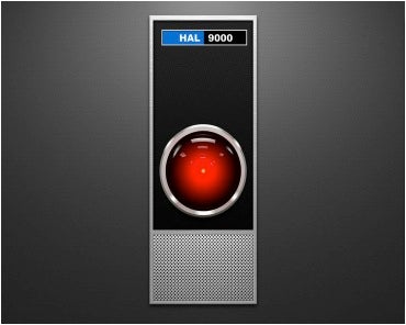 Will HBP name their simulated human brain computer HAL Jr.? HAL 9000 is a fictional character in Arthur C. Clarke's Space Odyssey series. The primary antagonist of 2001: A Space Odyssey, HAL (Heuristically programmed ALgorithmic computer) is a sentient computer (aka artificial intelligence) that controls the systems of the Discovery One spacecraft and interacts with the ship's astronaut crew. HAL's physical form is not depicted, though it is visually represented as a red television camera eye located on equipment panels throughout the ship (taken from Wikipedia and Bing Images).
According to Nature, many have raised doubts that HBP will achieve its goal, arguing that too little is understood about how the brain works to make the ambition feasible. Nevertheless, the Editorial adds, the project’s scope was originally conceived as a program whereby ‘bottom-up’ experimental data—electrophysiological, anatomical or cellular—would feed into a supercomputer without preconceived ideas of how the simulated neuronal circuitry might organize itself. A ‘top-down’ element has now been introduced.
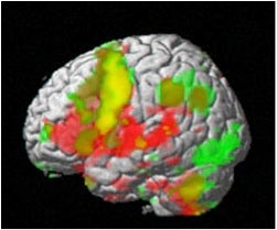 An fMRI of the brain. Green areas were active while subjects remembered information presented visually. Red areas were active while they remembered information presented aurally (by ear). Yellow areas were active for both types (taken from stanford.edu via Bing Images)
The ‘bottom-up’ data feed—mostly from research on mice—remains a core component, but how it is processed in the brain simulator will be guided by the findings of one of the Human Brain Project’s 15 subprojects, on high-level human cognitive architecture. This will generate data for both animals and humans, describing how cognitive tasks, such as those involving space, time and numbers, are processed in the brain. For example, in one major research project, around ten people will be selected for repeated study during the decade-long project. Their ‘reference brains’ will be measured using a range of non-invasive techniques such as functional magnetic resonance imaging (fMRI) and electroencephalography (EEG) to work out how the relevant neurocircuitry is organized during specific tasks. The detailed bottom-up data will have to align with this broad architecture.
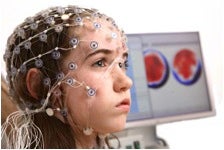 An EEG measures voltage fluctuations resulting from ionic current flows within the neurons of the brain. In clinical contexts, EEG refers to the recording of the brain's spontaneous electrical activity over a short period of time, usually 20–40 minutes (taken from buzz-master.com via Bing Images).
Supercomputing has proved too slow for real-time brain simulation, so other subprojects will focus on developing faster supercomputers, as well as neuromorphic computing, which can theoretically simulate brain activity orders of magnitude faster than occurs in a real brain.
HBP may still fail to deliver on its central promise, at least at the desired degree of sophistication, according to the Editorial, and it remains a high-risk initiative—keeping the unwieldy, multidisciplinary consortium of participants—mostly in Europe but also in countries including Israel, Japan and the United States—on track may also prove difficult. But the risks are spread over the subprojects, some of which will inevitably add significantly to our sum neuroscience knowledge.
The Editorial concludes by cautioning that, before getting too ‘starry-eyed about mega-projects, let’s remember that major breakthroughs in understanding the brain will continue to emerge from the labs of individual investigators. The journey towards a full understanding of the brain will be long and uncertain, and there will be ample opportunity for individual contributions to help point the way.’
I fully agree with this outlook, and believe it applies equally to the USA BRAIN Initiative.
Sex and brains: vive la difference!
 Ragini Verma, PhD, Associate Professor of Radiology, Department of Radiology (taken from academic. research.microsoft.com via Bing Images).
This is the catchy title of a Science and Technology article in the December 13th issue of The Economist about Prof. Ragini Verma’s PNAS publication about brain ‘hard wiring’ differences between the sexes pictured in my introduction above.
Before reiterating her conclusions, let’s briefly consider how these ‘hard wiring’ connections are experimentally determined. Dr. Verma’s technique is diffusion tensor imaging (DTI), which—according to the Miami Children’s Brain Institute—is a specific Magnetic Resonance Imaging (MRI) modality that measures local microstructural characteristics of water diffusion within tissues. As with other types of MRI, the data set is encoded upon geometric anatomical data. This property allows it to be used to create visual representations of the data, i.e. images derived from water diffusion throughout the brain. Because the fibers that connect nerve cells have fatty sheaths, the water in them can diffuse only along a fiber, not through the sheath. So, DTI is able to detect bundles of such fibers, and see how they’re connected—voilà!
While Verma’s DTI results showing sex-related ‘hard wiring’ differences are open to interpretation, her take according to Nature is that they underlie some of the variations in male and female cognitive skills. The left and right sides of the cerebrum, in particular, are believed to be specialized for logical and intuitive thought, respectively.
In her view, the wiring-diagram cross-talk between them in women, helps explain their better memories, social adeptness, and ability to multitask, all of which benefit from the hemispheres collaborating, so to speak.
In men, by contrast, within-hemisphere links let them focus on those things that do not need complex inputs from both hemispheres, hence the preoccupation with a single idea or thought. Because each half controls, by itself, only one half of the body, there is a physical basis for men having better motor abilities—or in common language, are better coordinated than women.
Dr. Verma’s other salient finding is that these connectome differences develop with age. The brains of boys and girls aged 8 to 13 demonstrated only few differences, but all of these differences became more pronounced at ages 13 to 17, and even more pronounced after 17 years of age. Differences in brains visible by DTI thus manifest themselves mainly when sex itself begins to matter.
By the way, it dawned on me that, based on Dr. Verma’s findings, the HBP’s ‘human brain in a computer’ could/should have two alternative modes of operation: male or female!
Closing Caveats
When researching Dr. Verma’s PNAS publication, I came across a Los Angeles Times account by Geoffrey Mohan that referred to researches who 'cautioned that the imagery is an indirect measure of axons, not a cell-by-cell census and map. And the results are strictly statistical averages, although in a very large sample.' Put another way, individual DTIs of other males and females may show significant deviations from these average patterns of connectivity.
Mohan’s article generated over 80 comments—some more interesting or humorous than others. I found that the following comment by 'craigbhill' on December 5, 2013 raised some additional caveats, mixed with some wit.
The key would be whether these gender-brain-based abilities cross all cultural bounds, then we would know they are innate. There's no denying the basic male-female global tendencies, it's not like females only in the West or even just in the US act thus and so when it's the bleepin' same everywhere, including in cultures removed from the broader worldwide culture. Sure, there are differences culturally, but not basically, and those changes are relatively minute within the basic natural behaviors.
So there's both nature and nurture, and we're now learning better than ever which and where.
(Accomplished by my basically male mind which has also been shown by a brain scan to act within the hemispheres in a more female way than most men's---I am blessed.)
Valentine’s Day, Love and fMRI
In the now classic 1984 track What’s Love Got To Do With It?, Tina Turner sings:
What's love got to do, got to do with it?
What's love but a second hand emotion?
You’ll have to read the other lyrics to find out her thoughts about that, but in the meantime, I’d like to connect the three topics of this section headings by first sharing what Wikipedia says about Valentine’s Day, partly because—honestly—I never knew its interesting history.
St. Valentine's Day—also known as Saint Valentine's Day, Valentine's Day or the Feast of Saint Valentine—is observed on Feb 14th each year in many countries around the World, and began as a liturgical celebration of one or more early Christian saints named Valentinus. The most popular martyrology associated with Saint Valentine was that he was imprisoned for performing weddings for soldiers who were forbidden to marry and for ministering to Christians, and were persecuted under the Roman Empire. During his imprisonment, he is said to have healed the daughter of his jailer. Legend states that before his execution he wrote her a letter signed "Your Valentine" as a farewell.
 Antique Valentine’s card (taken from Wikipedia).
The day was first associated with romantic love in the circle of Geoffrey Chaucer in the High Middle Ages, when the tradition of courtly love flourished. In 18th-century England, it evolved into an occasion in which lovers expressed their love for each other by presenting flowers, offering confectionery, and sending greeting cards (known as "valentines").
Ok, back to science. Obviously, love is an emotion, and I wondered whether fMRI has been applied to mapping brain patterns of love and other human emotions—none of which I regard to be ‘second hand’ as did Tina Turner. Somewhat to my surprise, this has indeed been done by fMRI. Here are some interesting—I think—findings posted on Psychcentral.com on June 21st 2013.
For the first time, scientists have identified which emotion a person is experiencing based on brain activity. A team at Carnegie Mellon University (CMU) lead by Karim Kassam, PhD, combined fMRI and machine learning to measure brain signals to read emotions in individuals. The findings illustrate how the brain categorizes feelings, giving researchers the first reliable process to analyze emotions.
Until now, research on emotions has been long stymied by the lack of reliable methods to evaluate them, mostly because people are often reluctant to honestly report their feelings. Further complicating matters is that many emotional responses may not be consciously experienced.
For the study, 10 actors were scanned at CMU’s Scientific Imaging & Brain Research Center while viewing the words of nine emotions: anger, disgust, envy, fear, happiness, lust, pride, sadness and shame.
While inside the fMRI scanner, the actors were instructed to enter each of these emotional states multiple times, in random order.
The computer model was able to correctly identify the emotional content of photos being viewed using the brain activity of the viewers.
The computer model achieved a rank accuracy of 0.84. Rank accuracy refers to the percentile rank of the correct emotion in an ordered list of the computer model guesses; random guessing would result in a rank accuracy of 0.50.
Next, the team took the machine learning analysis of the self-induced emotions to guess which emotion the subjects were experiencing when they were exposed to the disgusting photographs. The computer model achieved a rank accuracy of 0.91. With nine emotions to choose from, the model listed disgust as the most likely emotion 60 percent of the time and as one of its top two guesses 80 percent of the time.
Finally, they applied machine learning analysis of neural activation patterns from all but one of the participants to predict the emotions experienced by the hold-out participant.
This answers an important question: If we took a new individual, put them in the scanner and exposed them to an emotional stimulus, how accurately could we identify their emotional reaction? Here, the model achieved a rank accuracy of 0.71, once again well above the chance guessing level of 0.50.
“Despite manifest differences between people’s psychology, different people tend to neurally encode emotions in remarkably similar ways,” noted Amanda Markey, a graduate student in the Department of Social and Decision Sciences.
The research team also found that while on average the model ranked the correct emotion highest among its guesses, it was best at identifying happiness and least accurate in identifying envy. And, it was least likely to misidentify lust as any other emotion, suggesting that lust produces a pattern of neural activity that is distinct from all other emotional experiences.
Frankly, I did not do well in my college ‘Psychology 101’ course, I’m not sure what all of these fMRI results mean, but I suspect that several PhD theses will offer detailed interpretations. It’s nice to see that happiness was recognized the best. I do wonder, however, whether the results apply to non-actors?
As always, your comments are welcomed.
Postscript
After finishing the above post, the New York Times weekly Science Times section had a lengthy article by James Gorman entitled The Brain, in Exquisite Detail. The article featured state-of-the art NMR imaging by the Human Connectome Project, a $40 million 5-year consortium-effort supported by the NIH aimed at the “first interactive wiring diagram of the living, working human brain.”
To build this diagram, researchers are said to be “doing brain scans and cognitive, psychological, physical and genetic assessments of 1,200 volunteers. They are more than a third of the way through collecting information.” Data processing will yield a 3-D interactive map of the healthy human brain showing structure and function, with detail to one and a half cubic millimeters.
While this article is definitely worth a quick scan—pun intended—its closing remarks given below provide a sobering dose of reality in metaphorical terms.
“Perhaps the greatest challenge is that the brain functions and can be viewed at so many levels, from a detail of a synapse to brain regions trillions of times larger. There are electrical impulses to study, biochemistry, physical structure, networks at every level and between levels. And there are more than 40,000 scientists worldwide trying to figure it out.
This is not a case of an elephant examined by 40,000 blindfolded experts, each of whom comes to a different conclusion about what it is they are touching. Everyone knows the object of study is the brain. The difficulty of comprehending the brain may be more aptly compared to a poem by Wallace Stevens, “13 Ways of Looking at a Blackbird.”
Each way of looking, not looking, or just being in the presence of the blackbird reveals something about it, but only something. Each way of looking at the brain reveals ever more astonishing secrets, but the full and complete picture of the human brain is still out of reach.”